content-image
00

«Imagine»

They may not be a hit on Instagram yet. But the sharp pictures of proteins and small cellular structures taken by cryo-electron microscopes have gone viral in the scientific community. Their precision has already proven useful in the fight against three infectious diseases.

Text by Goran Mijuk and K.E.D. Coan, photos by Christian Jaggi

scroll-down
Home
en
de
zh
jp
Share
Share icon
content-image
Enter fullscreen

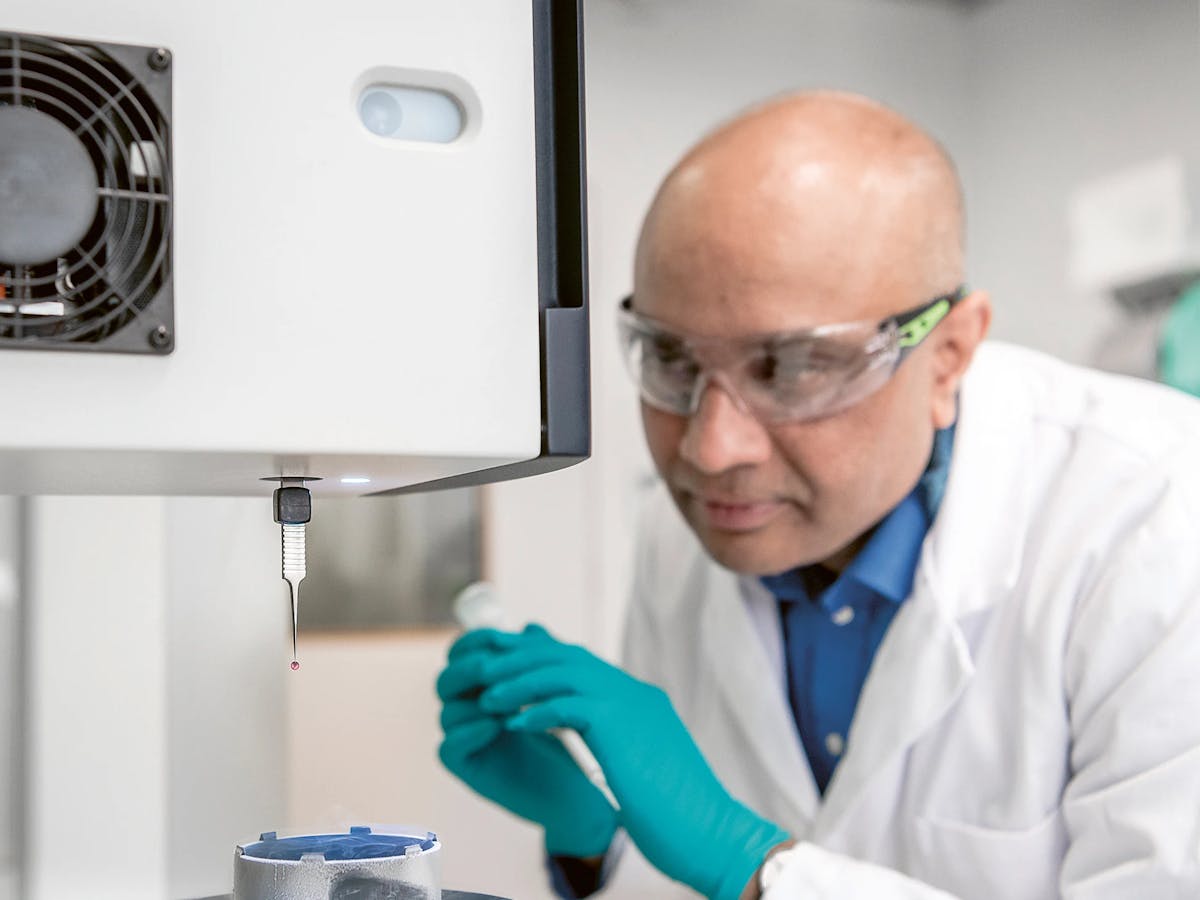
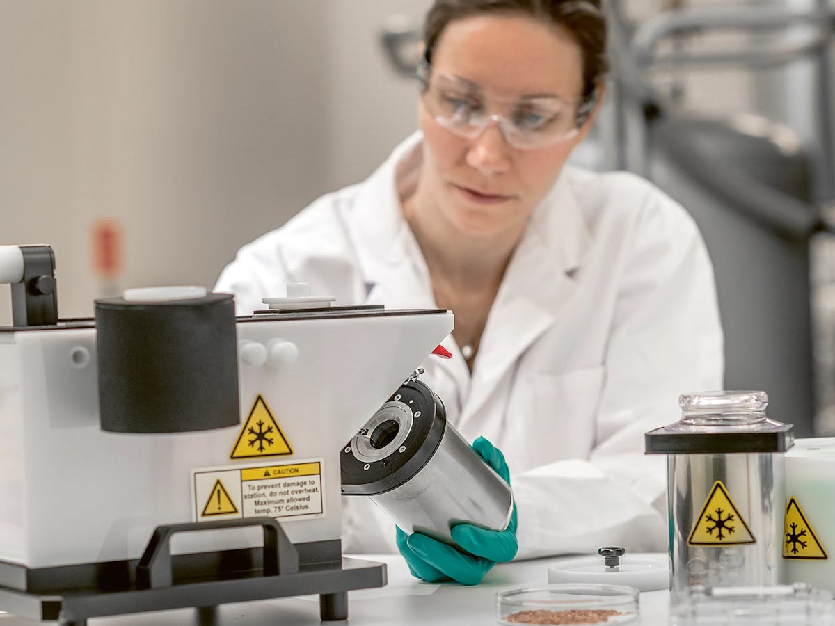
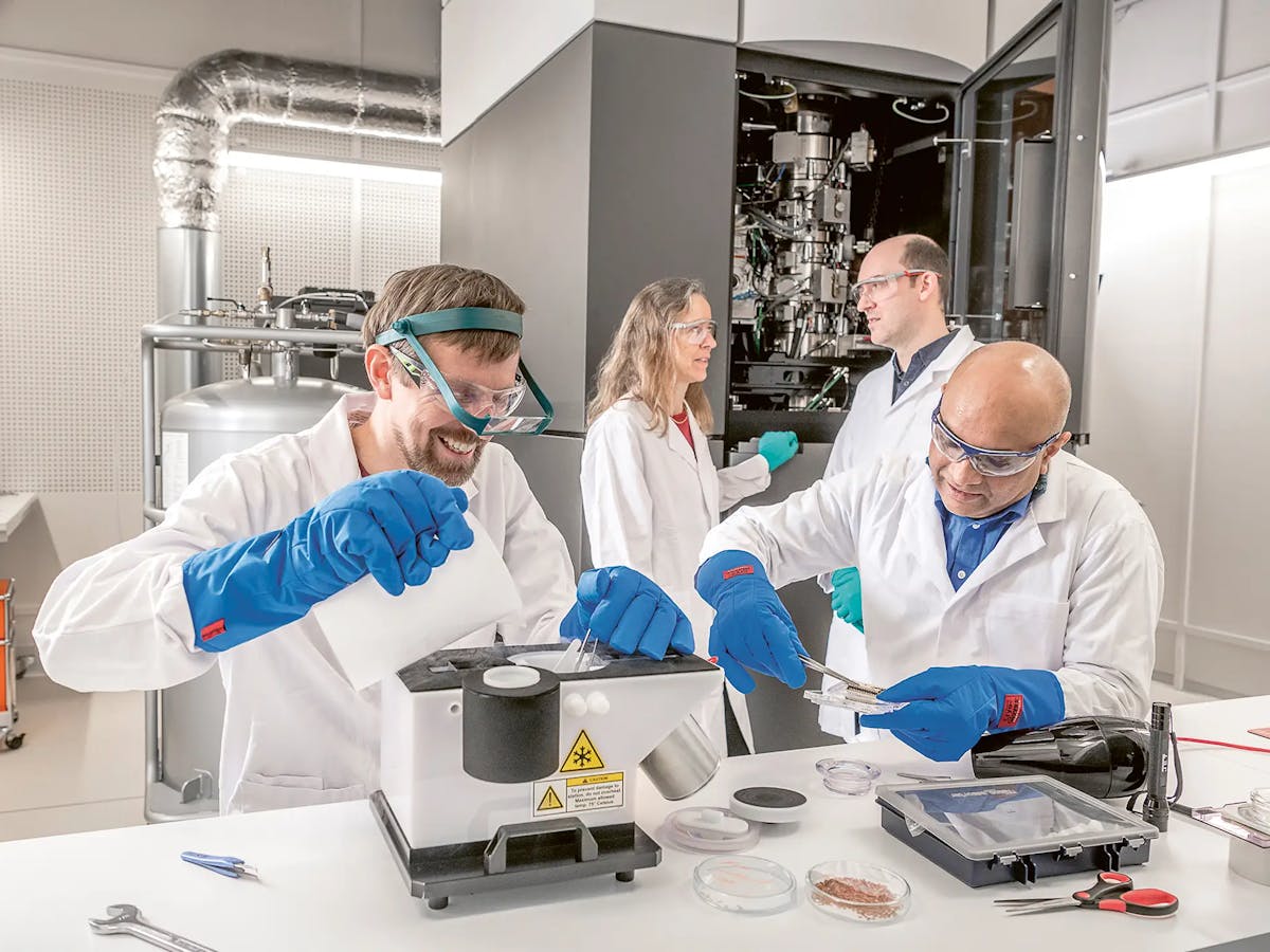
Researchers from Novartis and the FMI prepare a protein sample, which is later analyzed with the help of the cryo-electron microscope.

Published on 02/11/2020

It was a great day for science and a proud moment for Switzerland when Jacques Dubochet from the small town of Morges received the Nobel Prize in chemistry in 2017.

Together with Joachim Frank and Richard Henderson, with whom he shared the award, the retired professor from the University of Lausanne had provided the groundbreaking research that gave rise to cryo-electron microscopy.

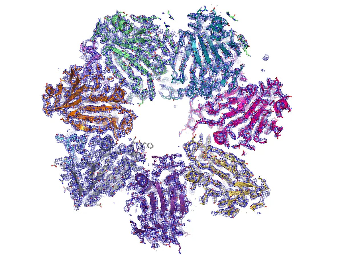
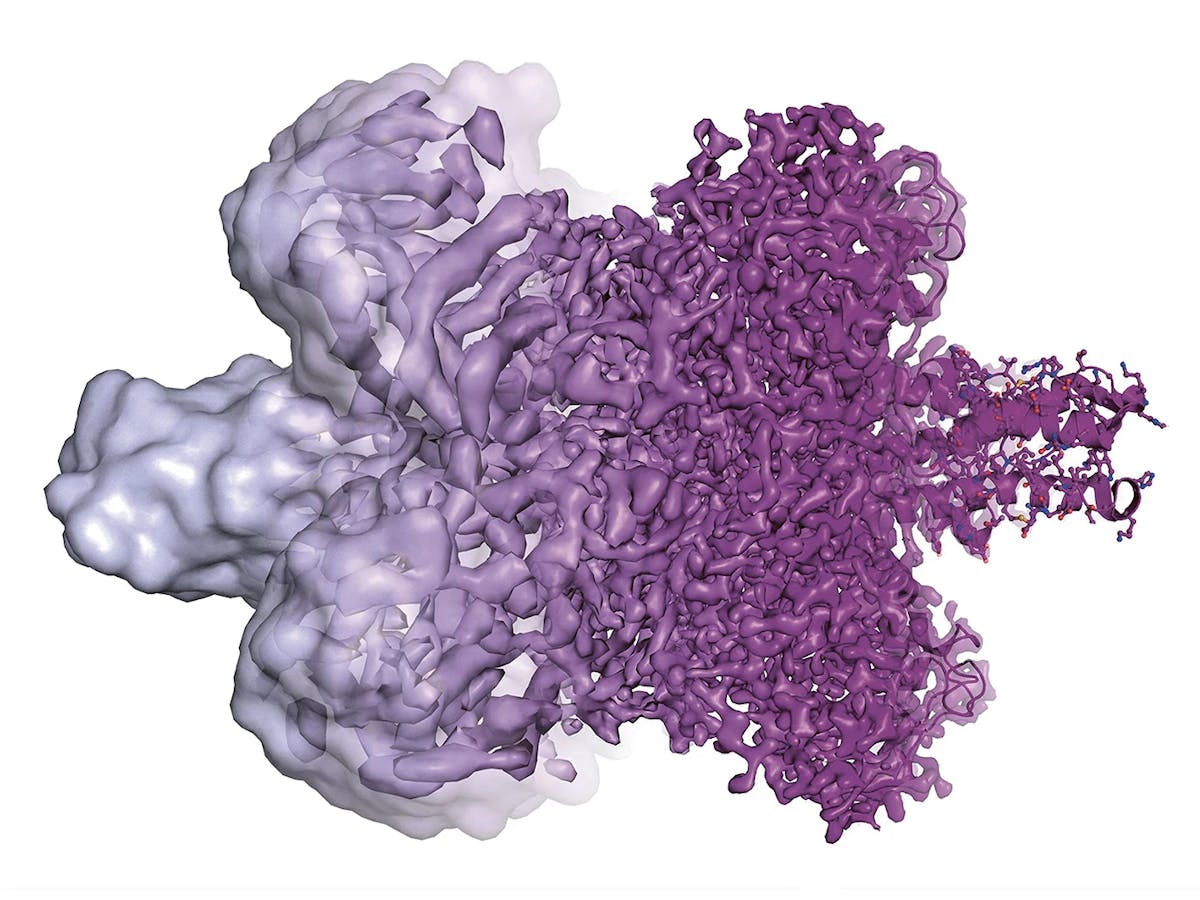
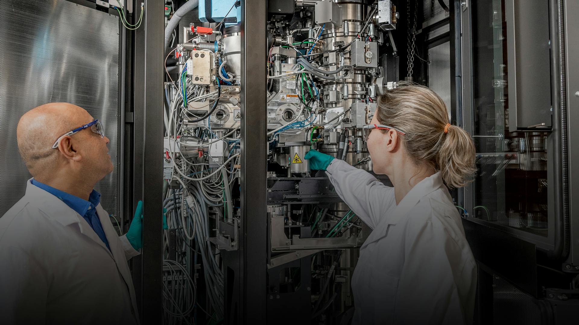
Cryo-EM, as it is known for short, is a new imaging technology which allows stunning views of complex molecular structures such as proteins.

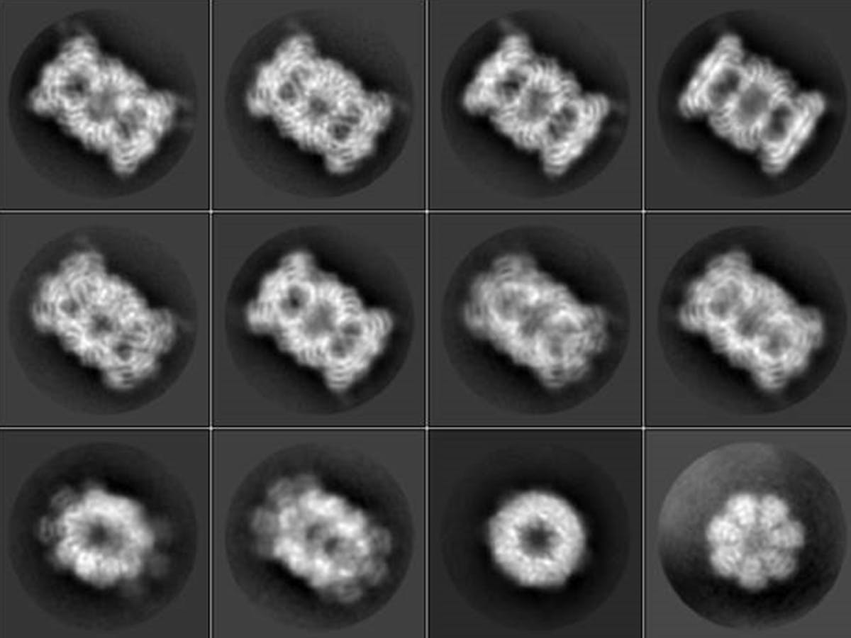
The Nobel Prize for Dubochet and his colleagues had been expected for a couple of years already because the image technology had leapfrogged to previously unknown resolution levels within a short period of time. Only 10 years ago, images of proteins developed with the help of a cryo-­electron microscope were blurred at best and were laughingly referred to by insiders as “blobs.” But then, rapid advances in digital technology and much improved cameras helped create 3D images of breathtaking sharpness, revealing nature’s beauty in hitherto unthinkable clarity.

Gold standard techniques such as protein crystallography and nuclear magnetic resonance spectroscopy almost paled in comparison.

Scientists around the world were aflame with excitement.

content-image
Enter fullscreen

Difference of resolution between traditional imaging technologies. The right-hand side shows the detailed structure of a protein rendered with the help of a cryo-electron microscope.

One of them was Nico Thomae from the Friedrich Miescher Institute for BioMedical Research in Basel. When he first saw one of these crystal clear 3D images in 2014, he was left speechless. He knew instantly that he wanted to work with this innovative tool, in which a protein sample is flash-frozen and then screened many times over with electrons to eventually produce a 3D image of a molecular complex.

“When I first saw what was becoming possible with cryo-electron microscopy, it blew me off my seat,” said Thomae. “It was clear that this was a revolution and that we were going to see a quantum leap in what was possible.”

Although Thomae had no doubts about the potential of cryo-EM, he had a few challenges to overcome before he could eventually work with such a machine.

Firstly, cryo-electron microscopes are far from cheap. Secondly, they are not easy to use. And thirdly, the value of the novel 3D image technology for medical research and drug discovery was not immediately obvious a few years back.

One of the researchers who saw the huge potential in the technology was Sandra Jacob, a structural biologist who works as an executive director of Protein Sciences at the Novartis Institutes for BioMedical Research in Basel.

When she was contacted by Thomae, the two of them worked toward convincing both Novartis and the FMI to provide financing for a facility in which researchers of both groups could work with the new technology. Such collaboration, they argued, could help them elucidate hitherto unknown biological structures and speed up the design of new compounds.

“The best groups in the world are all shifting to cryo-EM, but it’s too much of an investment to do alone – even for most pharmaceutical companies,” Jacob said. “But after Nicolas contacted us, we knew that we could learn from each other and that this was something that we should do together.”

Thumbnail
Play iconPlay icon
The beauty of science seen through the lense of a cryo-electron microsope
content-image
Enter fullscreen

Sample preparation is time-intensive...

Although they initially met with some skepticism, their efforts paid off. The collaboration between the two partners started already in 2015 and became operational a year later. Although it is just one of over 300 research partnerships maintained by Novartis, this one is special, says Sandra Jacob, because both partners share a common facility – a rarity in the industry.

This close co-habitation has already paid off, as the early results have exceeded all expectations: The team was able to produce high-resolution structures of biological complexes and has even made progress in the area of drug discovery thanks to the fact that the new technology also produces sharp images of assemblies of multiple proteins working together.

One such assembly is the so-called proteasome. This vital protein complex, which helps organisms degrade unwanted or misfolded proteins, had earlier been established as a potential drug target by the Genomics Institute of the Novartis Research Foundation.

GNF researchers, together with scientists from NIBR, had identified a single small molecule that can block the proteasome of three tropical parasites which cause leishmaniasis, Chagas disease and sleeping sickness.

Since the experimental compound does not affect the function of the human proteasome, researchers hope it may provide a way to selectively kill parasites that have invaded a human body.

Despite this knowledge, the scientists still lacked a clear picture of exactly how the compound killed the parasites’ proteasomes, groping in the dark as to how the compound could be developed into a drug.

Thumbnail
Play iconPlay icon
Cryo Podcast
content-image
Enter fullscreen

The cryo-electron team from left to right: Andreas Schenk, Christel Genoud, Srinivas Honnappa, Simone Cavadini, Sandra Jacob, Celine Be, Alexandra Graff, Nico Thomä, Chris Wiesmann.

This is where Christian Wiesmann, the director of the new cryo-EM center for Novartis, came in.

A researcher with many years of experience in X-ray crystallography, he and his team were able to establish the actual structure of the proteasome. This is now paving the way to improvements in the potential medical compound, which one day might help treat some of the deadliest tropical diseases. “There were a couple of proteasome models, but we couldn’t tell which of them was right – then, with cryo-EM, the answer was suddenly clear.”

Going forward, the team hopes that more such breakthroughs will be possible. Understanding disease pathways and seeing the actual structure of a disease-triggering cellular complex may help researchers develop compounds that snugly fit into these structures and stop a disease in its molecular tracks.

“Since the instrument arrived, we’ve had the feeling that we need to be even more ambitious, because what seemed next to impossible before, now seems very doable,” says Thomae. “These are door-opening moments in a scientist’s life, they don’t come often, and we’re really happy to be working as a team with Novartis to help drug discovery.”

Collaboration, meanwhile, will certainly play a crucial role going forward and may even take new forms.

At the end of his Nobel lecture, Jacques Dubochet made a plea to his research colleagues to imagine a world in which knowledge is shared in a new and open way.

Just as Dubochet used his fantasy to create stunning pictures of some of the world’s smallest things, he also urged his audience to think big and create a new vision of the world of science and healthcare, playing John Lennon’s song Imagine at the end of his lecture. Dubochet’s ideas may go viral one day as much as his images did.

icon

Home
en
de
zh
jp
Share
Share icon