
Published on 19/12/2022
“When you solve a protein structure, it’s like somebody switches on the light,” says Christian Wiesmann, who heads one of the most advanced microscopy labs in the world on the Novartis Campus in Basel. “If you’re lucky, you can suddenly understand how a whole biological system works,” Wiesmann explains.
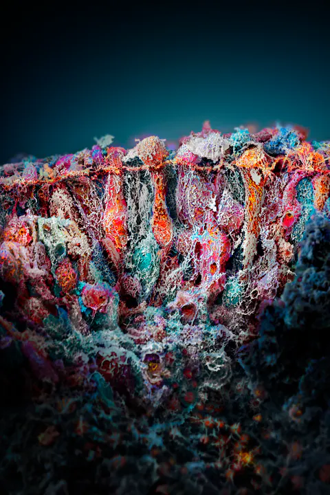
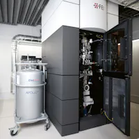
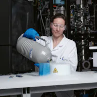
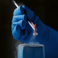
Much of his enthusiasm is down to the cryo-electron microscope, or cryo-EM, a Nobel Prize-winning imaging tool, which for laymen may look like an old-fashioned telephone booth with a maze of cables. The high-tech microscope, however, can render high-resolution images of proteins and help researchers better understand these biological building blocks of life whose understanding for medicine is instrumental. Through chemical biology, researchers can then develop custom-built chemical “tools” that fit in the pockets and grooves of protein surfaces – providing biological insights as well as starting points for new drugs.
Together with his team, which includes a handful of scientists from the Novartis Institutes for BioMedical Research, Wiesmann has been focusing on detecting the shape of highly complex and large proteins, an endeavor that, with traditional imaging tools, was considered almost impossible just a few years ago.
“Thanks to the cryo-EM, we now have access to so many important proteins which we couldn’t investigate with other methods. Since we started our facility in 2016, cryo-EM has already contributed greatly to our drug discovery efforts,” says Wiesmann.
A long path
Proteins, which are essential to most biological processes and can be the key cause of a disease if they malfunction, have long been puzzling scientists. For most of the early history of modern medicine, proteins were not even on the radar of drug researchers, who routinely tested the effects of medical compounds in cells, animals and patients, but lacked a molecular understanding of these structures and how they work in the body. Only with the development of X-ray technology, which was discovered around the turn of the 20th century, did scientists gradually learn about the importance of proteins and begin to understand more about their behavior, which allows them to fold in a fraction of a second into structures that are as mysterious as they are beautiful and vital to life on earth.
It took more than 50 years of hard work and a series of Nobel Prize-winning technologies before scientists were able to discern what proteins really look like and what their function is. One of the early breakthroughs was X-ray crystallography, for which researchers Max Perutz and John Kendrew won the Nobel Prize in 1962. The technology allowed scientists to study proteins in a crystalline form to get a sense of their shape and to study their function.
X-ray technology, however, needed years of fine-tuning before researchers could start to catalogue some of the nearly 30000 proteins that exist in the human body alone and the technique has remained fraught with challenges. “I started my professional life learning crystallography, and when I received my Ph.D. in 1996, solving just one crystal structure could take years and was sufficient to earn a Ph.D.,” says Wiesmann. “The biggest challenge is obtaining enough protein and to coerce the protein into forming crystals – and there are numerous examples of proteins that researchers have been trying to crystallize for decades without success.”

The rise of structural biology
Technological advances accelerated though and gave rise to a new field of drug development. “From the 1960s through the 1990s, most of our structural insights into the functioning of proteins and our understanding of disease mechanisms came from X-ray crystallography,” says Sandra Jacob, Executive Director at NIBR, who has been with Novartis since its formation 25 years ago and Ciba-Geigy before then.
Jacob heads NIBR’s structural biology group and was one of the leaders behind bringing cryo-EM to Novartis. “Then, around the year 2000, X-ray crystallography became much more powerful due to technical developments, and we began to use protein structures to think of specific ways of targeting these proteins and making more effective drugs.”
Once it was possible to truly understand the structure of these proteins – with their pockets and hinges – the field could literally see where drugs bind to their protein targets and how that changed their activities. This gave rise to tailor-made drugs designed to fit exactly in the right spots to stop or control their protein targets.
The field of chemical biology – which brings together the power of structural biology and chemistry – is rooted in these technical developments as well.
“That was a turning point across the entire pharmaceutical industry,” says Jacob. “After we proved that structural tools could really make a difference in drug discovery, our structural biologists were overwhelmed with requests from drug discovery teams and they really focused on getting us involved early in the process.”

Innovation-minded and partnerships
Within a short time, Novartis was not only able to prove that the structural analysis of proteins could help drug developers create targeted therapies. The teams were also able to bring several compounds to the market, creating new drug development processes that would usher in what today is known as personalized medicine.
That Novartis would be at the forefront of this development is mainly due to its unrelenting commitment to medical innovation. Ever since the company was founded in 1996 through the merger of Ciba-Geigy and Sandoz, Novartis has been pushing medical boundaries, extending a tradition that started more than 100 years ago when Sandoz and Ciba-Geigy launched their medical research.
While investments stood at about 2 billion US dollars at the time of the merger, annual research and development investments were continually increased and now amount to around 9 billion dollars every year, giving rise to one of the world’s most powerful development pipelines with more than 200 ongoing clinical trials.
During this time, Novartis not only developed several breakthrough therapies, it also built one of the most powerful research institutes. NIBR today is home to more than 5000 scientists who work together with colleagues from the Novartis clinical development and academia such as the Paul Scherrer Institute, which is a key partner in the area of X-ray crystallography.
“We have a lot of connections and contacts with the academic world related to advanced technologies,” says Sandra Jacob. “We’ve been an early partner of academia and have always worked with some of the best partners in the industry, also to get hold of the most advanced technologies to push our knowledge and expertise.”

New frontiers
This is also the case with cryo-EM for which Novartis is partnering with the FMI, which also has close ties to academic research. Moreover, the technology is opening up new possibilities for protein and drug research that is out of reach for traditional imaging technologies. “Cryo-EM is a rising star among structural biology methods because it’s opening doors for so many larger drug targets and protein complexes that cannot be crystallized and so have been out of reach until now,” says Wiesmann.
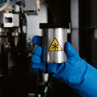
Because cryo-EM uses a beam of electrons to produce images of biological molecules frozen in a thin layer of ice, it effectively has no upper size limit. The technology has been slowly developing but witnessed a revolution in 2013, when improvements in hardware allowed the resolution achieved by cryo-EM to rival the one obtained with crystallography. Since then, the number of structures produced by cryo-EM has doubled every two years. In 2020, not even 10 years after the resolution revolution, more than 20 percent of new protein structures were determined by cryo-EMs.
“There are projections that within four years cryo-EM will become the most dominant structural biology method out there,” says Wiesmann. “It is amazing how rapidly this method has changed structural biology and how profoundly it is impacting our drug discovery efforts already.”
Beyond larger proteins, another field-changing advantage of cryo-EM is that researchers can now look at ensembles of proteins together – which was nearly impossible in the past. Proteins frequently act in coordination with other proteins, and cryo-EM is giving some of the first snapshots of how these proteins interact with each other.
The team has already captured structures of previously inaccessible drug targets for cancer and immune diseases. One of their targets, the proteasome, is an assembly of proteins that rids the cell of other unwanted proteins. The proteasome is also a potential drug target for several parasitic diseases like leishmaniosis, a disfiguring and potentially fatal disease that causes up to 30000 deaths each year. Cryo-EM has already helped the team identify and visualize a new molecule that kills the parasite by binding to the proteasome.
But Wiesmann, Jacob and their colleagues are not stopping here and are on the lookout for new frontiers. Alongside the most recent advances, there is one more method that Wiesmann and Jacob are looking forward to – methods that allow them to see proteins in action. “In drug discovery, all of these techniques come together and provide complementary information,” says Jacob. “Another real breakthrough for us will be methods that can actually see what’s happening to a protein in the cell – and we might see these tools in the near future too.”
Given the determination of Novartis to continue to boost innovation, new findings may soon pop up as researchers get a clearer image of nature’s tiniest structures.
You may also be interested in the following:


Thanks for reading Live.Magazine
Stay connected by subscribing to our newsletter
SubscribeBy submitting your email, you consent to Novartis AG collecting and processing your email data for Novartis internal use, in accordance with our privacy policy, and by protected technical means.