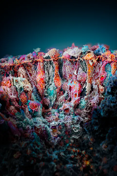
Magdalena Renner was skeptical and frustrated. For months she had been trying to produce retinal organoids in high enough quantities that would eventually help spur the basic research efforts at the Institute of Molecular and Clinical Ophthalmology Basel (IOB). But things did not turn out as she had wished. While she and her team were able to produce high-quality organoids that allowed scientists to study the function and disease genesis with the help of these artificial photoreceptor tissues, the number of organoids her lab could create reached a maximum of several dozen. This, she knew, was too little to make a difference and support scientists in being less dependent on difficult-to-acquire human retinas, as well as mouse retinas, which are not perfect research organisms in ophthalmology because they differ in important physiological traits. Unlike human eyes, for example, mice have no fovea, which is responsible for sharp vision. She needed to change something but was unsure what. Yet, she had a hunch, which would not only help boost production of these important research samples, but also unleash the IOB’s ability to do basic research at scale. Together with her colleagues, she was also able to pave the way to start the first large compound screening with retina organoids.
Magdalena Renner.
Organoid pioneer Renner was well acquainted with organoid research. She had already focused on this emerging field early on in her career when she joined the lab of Juergen Knoblich at the Institute of Molecular Biotechnology in Vienna, which paved the way in this domain by induced pluripotent stem cell technology in neurology more than a decade ago. Among other things, she was a co-author of a seminal paper that detailed the development process of such minibrains in 2013. In parallel, while doing her Ph.D. in Austria’s capital, she was also one of the first scientists to work on the question on how to use minibrains as disease models. After her stint in Vienna, where she also worked with emerging gene technologies such as CRISPR/Cas9 that allows for precision gene manipulation, Renner joined Novartis and the Friedrich Miescher Institute. There, IOB co-founder and today’s director Botond Roska asked her in 2017 to join the freshly founded IOB to work on retinal organoids – a little-explored field back then which needed to be set up from scratch. “The retina is part of the brain,” Renner said. “So, for me, the switch from brain organoids to retinal organoids was not huge. There are many parallels in the protocols, and the challenges are similar.”
Step by step As part of the production process involving induced pluripotent stem cells, Renner would take human cells and revert them back into stem cells – a cell type that can develop into any other cell type of the body. From there, she would try to develop them into retina organoids. As part of this technology, skin or blood cells are reprogrammed into an embryonic-like state. During this reversal process, the cells maintain a donor’s genome. This allows researchers to later study either healthy or diseased organoids. They can also genetically design organoids with specific mutations to better understand a biological process or an aspect of a disease. After the transformation into stem cells, Renner would initiate the growth process by putting the cells on small plates, add a cell culture medium and wait until the retinas became visible under the microscope – showing a horseshoe-like structure. Renner would then use a needle to scoop them out of the plate and transfer them to another nutrient vessel, where they would form into full organoids. As was industry standard back then, Renner would only take those organoids out of the plate that fit the horseshoe shape and discard the others. The net result of this process was that six months down the road, she and her team would only count a few retinas in the growth vessel. This was too little to make a big splash or even consider a compound screen to test a treatment or understand the genetic makeup of an organoid or cell type.
Following a hunch “The process takes a very long time, which is frustrating,” Renner said. While she was struggling to accelerate it, she was also questioning the established development protocol. “What made me suspicious was that there were no good criteria as to which organoids are fit for further processing,” she explained. One day, she decided, instead of painstakingly fishing out the horseshoe-shaped organoids, she would dissect all the organoids that were on the plate and transfer them to the nutrient vessel. “Instead of individually cutting off those structures from the cell culture dish, I took a pipette, drew a grid with it and scraped off all the cells on my dish, without worrying about what they looked like,” Renner said. The intuition to follow this route was not only due to the fact that she was not fully convinced by the established method as it lacked clear criteria beyond the organoids’ form. She had in fact observed that when she was taking out the horseshoe organoids – a process that often took days – organoids on the plate would form where she did not expect it. She took a leap of faith and decided to test a new procedure. “I was going to discard the plate anyway. So, in a sense, it cost me nothing to just scrape off everything and see what was there. To my astonishment, we suddenly went from 20 organoids to hundreds in almost no time.” The serendipitous moment took her by surprise. And while she concedes that much of the organoid development process remains in the dark and that even scientists marvel at the fact that induced pluripotent stem cells can develop into miniature organs, her luck was based on a scientific decision to test another route as the established one seemed ineffective. The move not only paid high dividends. It also showed that the IOB, just shortly after its inception, lived up to its ambition to attract leading talents who were willing to transform ophthalmology and change the status quo.
High quality Meanwhile, the quality of the organoids Renner’s team produced was also high. “We compared them to the human adult retinas. And the cells, especially cone photoreceptors, were amazingly similar,” Renner said. Although organoids are minuscule compared to real organs, in order to be efficient, they need to include the vital parts that are important to study. This helps scientists deepen their understanding of the underlying biology. Furthermore, they can also use the organoids to do genetic testing, which has improved rapidly since the advent of precision gene technology. Induced pluripotent stem cell technology and CRISPR/Cas9 gene technology have been a boon for science, helping researchers reduce animal testing and stay off controversial stem cell research. Besides minibrains and minikidneys, scientists are working on a series of new types of organoids around the world. For Renner, organoids have a huge capacity to spur scientific research if the quality of the organoids meets the necessary criteria. “In our organoids, there are five layers resembling those found in the adult human retina, including three nuclear layers where the retinal cell bodies reside, and two synaptic layers facilitating cell communication,” Renner said. Given the high quality and functional resemblance to the human retina, researchers now readily switch to organoids and have even embarked on large compound screens. One of the first such effort was a project led by Stefan Spirig from the IOB, who tested about 3,000 compounds with about 20,000 organoids from Renner’s lab. Other scientists at the IOB are following suit.
New horizons For Renner and her colleagues, the work continues, as they are now focusing on new model organisms such at retinal pigment epithelium, an important cell type that the team is trying to grow in a very pure way. “These are crucial cell types for interaction and play an important role in disease. To have them as organoids is important.” The team has succeeded in developing a new protocol that allows the generation of such cells to be used in high-throughput screens, much like in the case of the retina organoids that have been used by Spirig. The team has also developed a method to determine whether these cells can be used as a gene therapy prescreening tool to select the best candidates for validation in human explant cultures, which are difficult to obtain. “We try many things in parallel,” Renner said, “to develop model organisms that can help scientists reduce their dependency on both the human retina and the mouse retina. The overall goal is to build a better model of the eye and thus help researchers even more to advance our understanding of ophthalmology.”
Thanks for reading Live.Magazine
Stay connected by subscribing to our newsletter
SubscribeBy submitting your email, you consent to Novartis AG collecting and processing your email data for Novartis internal use, in accordance with our privacy policy, and by protected technical means.